Unit 2: Functional Diagnostics and Imaging Techniques
| Sitio: | iLearn - Lernmanagementsystem der Hochschule Deggendorf |
| Curso: | vhb Demo: Application of Medical Technology |
| Libro: | Unit 2: Functional Diagnostics and Imaging Techniques |
| Imprimido por: | Invitado |
| Día: | domingo, 21 de junio de 2026, 17:45 |
Learning Objectives
Upon completion of this unit, participants will be able to:
- Learn about medical imaging techniques.
- Identify the main benefits and risks of X-ray.
- Understand the main diseases and diagnostic tools utilized in the cardiology, gastroenterology and pneumology departments.
- Interpret the medical terminologies of functional diagnostics techniques.
- Understand the importance of using technology in various healthcare departments.
2.4 Pneumology
Section 1: Introduction to Pneumology
- What is Pneumology?
Pneumology is a medical specialisation which covers the conditions affecting the upper and lower respiratory system.
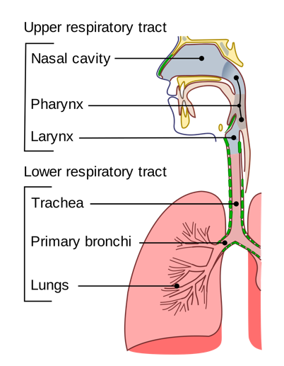
Figure 1: represents the lower and upper respiratory tract parts.
Source: AGOVIRAX; viral respiratory infections.
- What are the main functions of the respiratory system?
2.3 Pneumology
Section 1: Introduction to Pneumology
- What are the main functions of the respiratory system?
During inhalation, air containing a high concentration of oxygen enters the lungs through the airways. At the same time, blood is returning to the lungs with high carbon dioxide and low oxygen levels. In the pulmonary capillaries, carbon dioxide is exchanged for oxygen from the alveoli. Blood then leaves the lungs with a high oxygen content and a relatively low carbon dioxide content to be distributed to the tissues of the body by the left side of the heart. During exhalation, gas with a high concentration of carbon dioxide is expelled from the body.
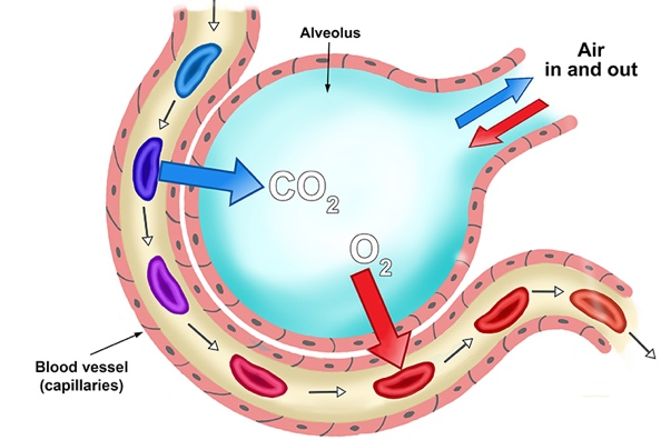
Figure 2: The alveolus is a sac which allows gas exchange of oxygen and carbon dioxide by diffusion.
The alveoli have thin walls to aid this diffusion.
Source: MAMMOTHMEMORY; the pulmonary system.
2.3 Pneumology
Section 1: Introduction to Pneumology
- What are the main functions of the respiratory system?
Excess carbon dioxide in the blood from breathing reacts with water to produce carbonic acid. This lowers the blood pH and increases the depth of respiration as shown in the following reaction:
CO2 + H2O ⇌ H2CO3 ⇌ H+ + HCO3-
The respiratory system can therefore participate in acid-base balance by removing CO2 from the body. The central nervous system has sensors for CO2 and hydrogen ion levels (H+) in the arterial blood and in the cerebrospinal fluid that send information to the body's controllers of breathing.
2.3 Pneumology
Section 1: Introduction to Pneumology
- What are the main functions of the respiratory system?
Phonation is the production of sounds by the movement of air through the vocal cords. Speech, singing, and other sounds are produced by the actions of the central nervous controllers on the muscles of respiration, causing air to flow through the vocal cords and the mouth.
2.3 Pneumology
Section 1: Introduction to Pneumology
- What are the main functions of the respiratory system?
The lungs tend to protect against the atmosphere environment microorganisms such as bacteria, dust, toxic gases and smoke (e.g. campfire smoke or tobacco smoke).
2.3 Pneumology
Section 1: Introduction to Pneumology
- What are the main functions of the respiratory system?
- a. Gas exchange:
- b. Acid-Base Balance:
- c. Phonation:
- d. Pulmonary defense mechanisms:
- e. Pulmonary metabolism and the handling of the bioactive materials:
The cells of the lung metabolize substrates to supply nutrients and energy for their own maintenance. The pulmonary capillary's endothelium contains a great number of enzymes that can produce, metabolize or modify naturally occurring vasoactive substances.
g. Functional diagnostics
1. Spirometry:
According to the United Kingdom's National Health System (NHS), spirometry is a simple test used to help diagnose and monitor certain lung conditions by measuring how much air a person can breathe out in one forced breath. It's carried out using a device called a spirometer, which is a small machine attached by a cable to a mouthpiece.
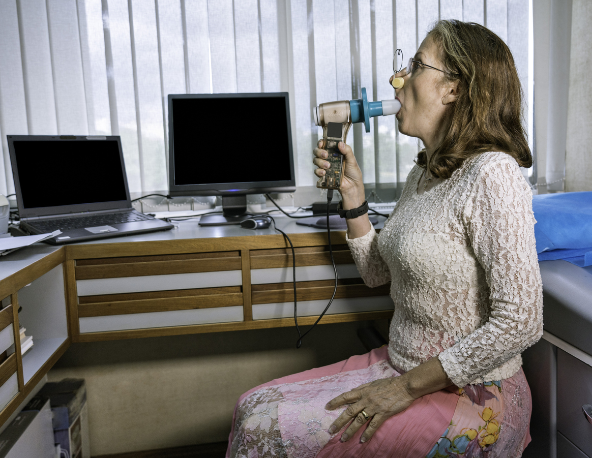
Picture source: (Link)
2. Finger Pulse Oximeter:
According to the UK's NHS, pulse oximetry is a noninvasive and painless test that measures oxygen saturation level. Oxygen saturation level is the amount of oxygen present in the blood. It can rapidly detect even small changes in how efficiently oxygen is being carried to the extremities furthest from the heart. The pulse oximeter is a small, clip-like device that attaches to a body part, like toes or an earlobe. It’s most commonly put on a finger and used in critical care settings like emergency rooms or hospitals. It is used to monitor the following conditions:
- Chronic obstructive pulmonary disease (COPD)
- Asthma
- Pneumonia
- Lung cancer
- Congenital heart defects
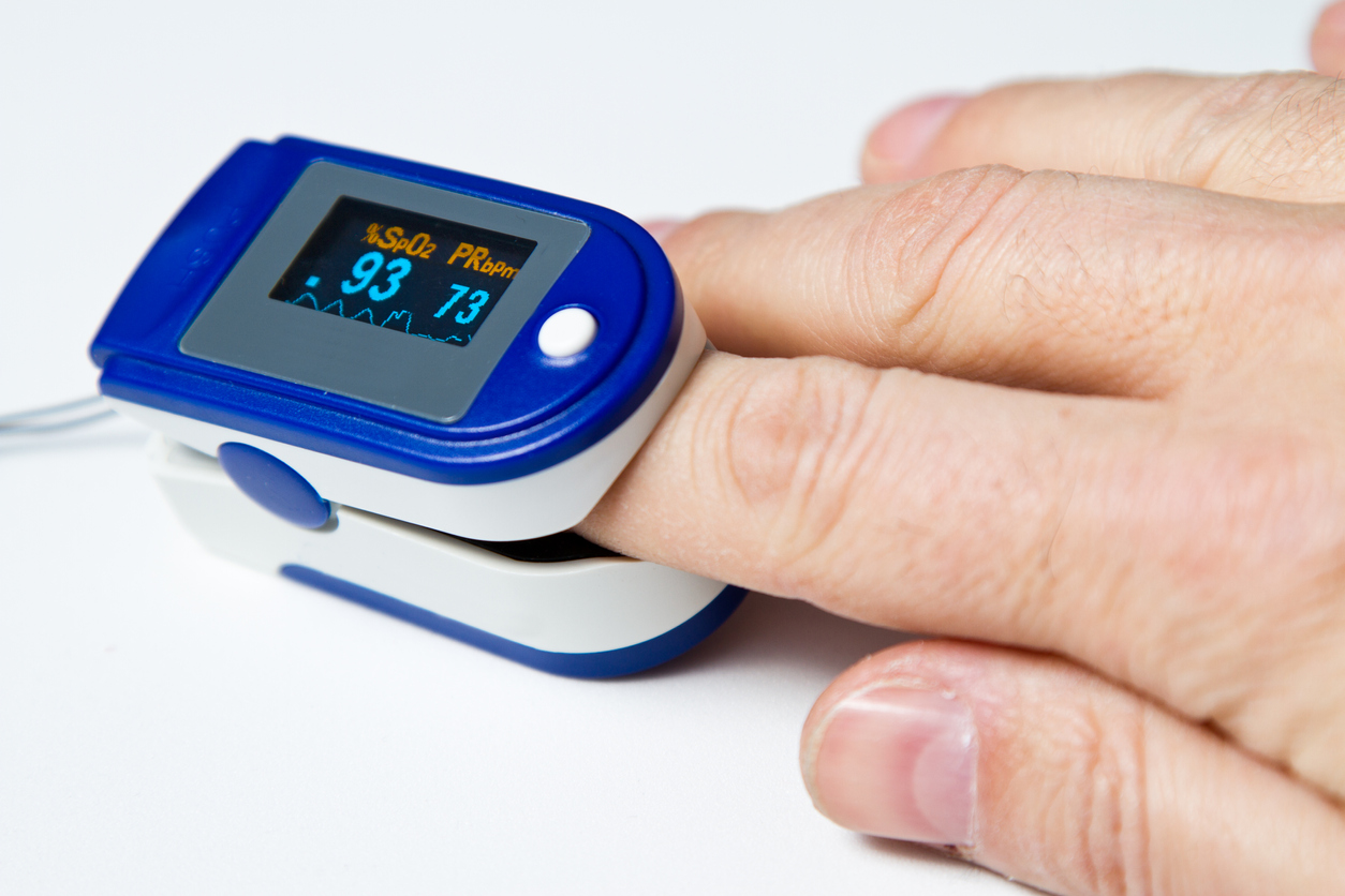
Picture source: (Link)
Lung problems like pneumonia and respiratory failure can be some of the most severe symptoms of the novel Coronavirus (the cause of COVID -19). Some individuals are prescribed a pulse oximeter for conditions that cause them to have periods of low oxygen, for certain underlying lung conditions, if someone with a lung condition is exercising or even traveling to high altitudes.
3. Bronchoscopy:
According to the NHS, a bronchoscopy is a procedure that allows a doctor to examine the inside of the lungs, including the bronchi. The bronchi are the main pathways into the lungs. During a bronchoscopy, a doctor inserts a thin tube containing a light and camera into the lungs through the nose or mouth. The doctor can use the findings to diagnose infections, tumors, or diseases in the lung.
There are two types of bronchoscopes that may be used: Flexible and rigid.
- Flexible bronchoscopy uses a long, thin, flexible lighted tube that can access the small airway branches. A sample of tissue or a biopsy may be obtained through the bronchoscope for laboratory study.
- Rigid bronchoscopy involves the use of a rigid, straight, hollow metal tube. Rigid bronchoscopy is utilized when the airway is obstructed by blood or foreign objects.
4. MRI and X-ray.
Further reading
The following is a list of further readings that you may find useful to enhance your understanding of the topics in this module:
Imaging Techniques:
- A Comparative Study of Medical Imaging Techniques
- How to reduce exposure to medical imaging risks?
- Benefit and risk of X-ray diagnostics
- MRI vs. X-ray
Cardiology:
Pneumology:
* You will be forwarded to an external page. We are not responsible for the content of this site. There may be other regulations regarding data protection.
References
- Bender J., Russell, K., Rosenfeld, L. & Chaudry, S. (2010). Oxford American Handbooks of Cardiology, Oxford American Handbooks of Medicine, 1st Edition.
- Douglas O. Faigel, MD, FACG, FASGE, Michael L. Kochman, MD, FACP & FASGE. (2007). Endoscopic Oncology; Gastrointestinal Endoscopy and Cancer Management, Human Press, Totowa, New Jersy.
- DRUGS, https://www.drugs.com/cg/upper-endoscopy.html.
- Hoyt R.E. & Yoshihashi A. (2014). Health Informatics: Practical Guide for Healthcare and Information Technology Professionals. 6th
- Keshav S. & Culver E. (2011). Gastroenterology; Clinical Cases Uncovered (CCU), WILEY- BLACKWELL, A Jone Wiley & Sons, Ltd., Publication.
- Keucherl M., Hagenmüller F. & Tajiri H. (2015). Video Capsule Endoscopy; A reference Guide and Atlas, Springer-Verlag Berlin heidelberg.
- Loscalzo J. (2010). Pulmonary and Critical Care Medicine. Derived from Harrison’s Principles of Internal Medicine, 17th Edition.
- Levitzky M. G. (2013). Pulmonary Physiology. Derived from MC Graw Hill Education, LANGE, 8th Edition.
- NIDDK, https://www.niddk.nih.gov/health-information/digestive-diseases/digestive-system-how-it-works.
-
Nelson R. & Staggers N. (2018). Health Informatics: An Inter- professional Approach. Elsevier, St. Louis, USA.
- YPO, https://www.ypo.education/medical-tests/barium-swallow-t311/video/.
- Venot A., Burgun A. & Quantin C. (2014). Medical Informatics, e- Health. Springer, Paris/Heidelberg.